Pyoderma Gangrenosum

Alison A. Smith, MD, PhD
, Frank H. Lau, MD

History:

A 29-year-old man presented with a three-year history of a painful and recurrent left posterior calf wound. The patient had a past medical history of Factor V Leiden, chronic bilateral lower extremity deep vein thromboses, and HIV. The patient was never a smoker, but did use marijuana. When the wound first appeared in 2015, it was healed with local wound care. However, in 2017, there was a spontaneous recurrence of the wound that occurred while wearing compression stockings.

29 yo man with a 3-year history of a recurrent left posterior calf wound measuring 9x7cm.

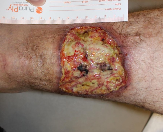
Fig.1. 29 yo man with a 3-year history of a recurrent left posterior calf wound measuring 9x7cm.

Findings:

Left posterior/distal calf wound measuring 9 x 7 cm. The wound had erythematous undermined edges and an ulcerated center with fibrinous exudate. No bone or tendon exposure was found.

A 29-year-old man with a recurrent left posterior  calf wound immediately post-debridement. Tissue biopsy of the wound revealed mixed pyoderma gangrenosum and vasculitis.

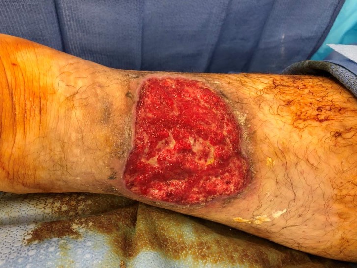
Fig.2. A 29-year-old man with a recurrent left posterior  calf wound immediately post-debridement. Tissue biopsy of the wound revealed mixed pyoderma gangrenosum and vasculitis.

Intra-operative application of dHACM to a lower leg wound of a 29 year old man with pyoderma gangrenosum.

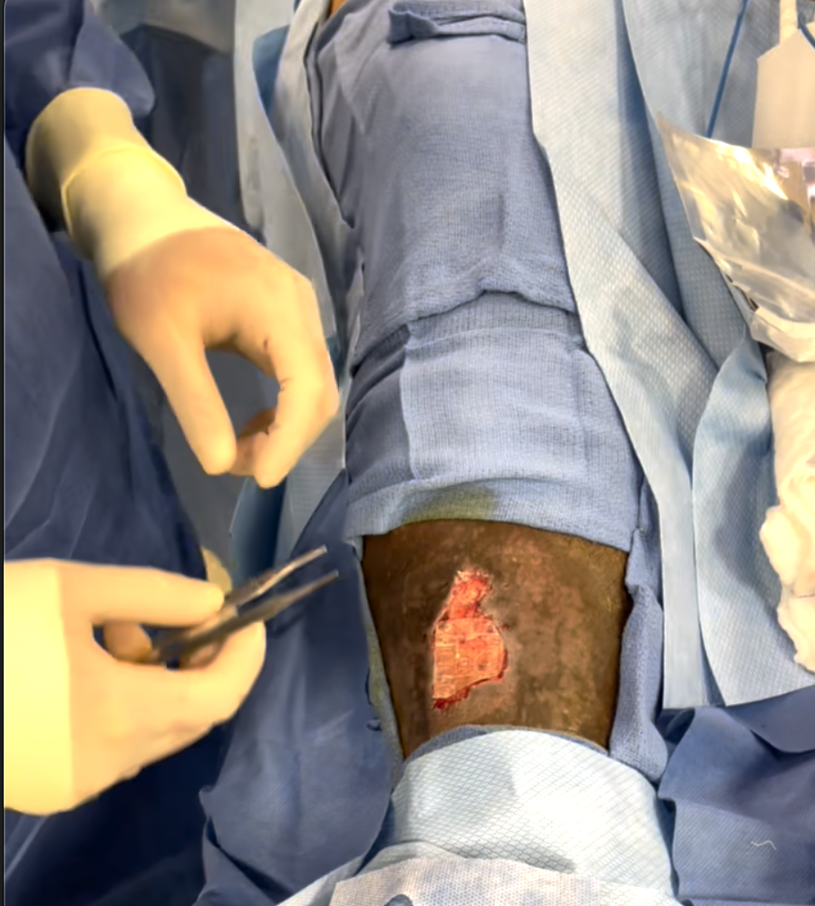
Fig.3. Intra-operative application of dehydrated human amnion/chorion membrane to a lower leg wound of a 29 year old man with pyoderma gangrenosum.

Differential Diagnoses:

Arterial insufficiency, venous stasis, infection, malignancy, vasculitis, trauma, cutaneous manifestations of other inflammatory disorders.

Workup Required:

Pyoderma gangrenosum is a diagnosis of exclusion. Routine workup included complete blood count, inflammatory markers erythrocyte sedimentation rate (ESR) and C-reactive protein (CRP), skin biopsy, and wound cultures. Other studies included doppler venous and arterial ultrasounds, liver and renal function tests, hepatitis screen, vasculitis, and hypercoagulability workups. Colonoscopy was considered but deemed not necessary in the absence of any signs of malignancy.

Plan:

Optimize wound care, avoid surgical trauma, manage pain adequately, and choose effective medical management to reduce the aberrant inflammatory response.

Expertise Needed:

Dermatologist and wound care specialist.

Treatment:

Cleansing with antibacterial agents and enzymatic/autolytic debridement were done. Sharp surgical debridement was avoided due to pathergy concerns. Dressings were selected to maintain a moist wound environment and to decrease local edema. First-line therapy involved topical or oral (0.5-1mg/kg/day) corticosteroids. Cyclosporine and TNF inhibitors were kept in mind as second and third-line therapies. Topical sodium cromoglycate, nicotine, 5-aminosalicylic acid, dapsone and oral methotrexate, mycophenolate mofetil, sulfasalazine, and azathioprine were considered for treatment. Oral acetaminophen, non-steroidal anti-inflammatory drugs, and opiates were used for pain management.
29-year-old man with a recurrent left lower extremity wound, diagnosed with pyoderma gangrenosum. Status post 1-week of treatment with dehydrated human amnion/chorion membrane (dHACM). The patient went on to receive a split-thickness skin graft.

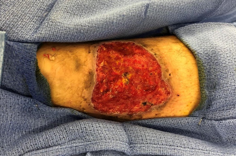
Fig.4. 29-year-old man with a recurrent left lower extremity wound, diagnosed with pyoderma gangrenosum. Status post 1-week of treatment with dehydrated human amnion/chorion membrane (dHACM). The patient went on to receive a split-thickness skin graft.

Fig.5. 29-year-old man with a recurrent left lower extremity wound, diagnosed with pyoderma gangrenosum. Status post 1-week of treatment with dehydrated human amnion/chorion membrane (dHACM). This is an image 5 days post-op from dHACM placement and after application of a split-thickness skin graft.

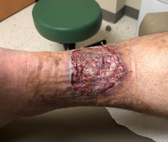
Fig.6. 29-year-old man with a recurrent left lower extremity wound, diagnosed with pyoderma gangrenosum. Status post 12 days of treatment with dehydrated human amnion/chorion membrane (dHACM) and split-thickness skin graft.


Follow Up:

The patient returned to clinic for follow up. On post-op day 21, he had evidence of good wound healing with incorporation of the graft.

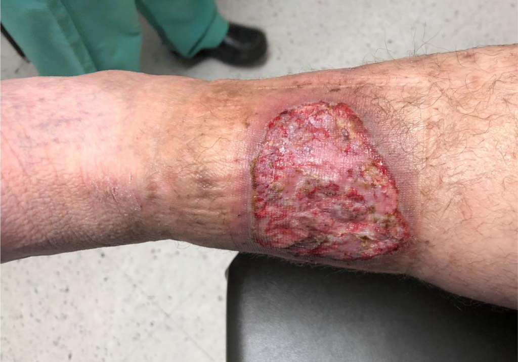
Fig.7. 29-year-old man with a recurrent left lower extremity wound, diagnosed with pyoderma gangrenosum. Status post 21 days of treatment with dehydrated human amnion/chorion membrane  and split-thickness skin graft.


Print Friendly, PDF & Email

References

Alavi A, French LE, Davis MD, Brassard A, Kirsner RS. Pyoderma Gangrenosum: An Update on Pathophysiology, Diagnosis and Treatment. Am J Clin Dermatol. 2017;18(3):355-372.
Ahronowitz I, Harp J, Shinkai K. Etiology and management of pyoderma gangrenosum: a comprehensive review. Am J Clin Dermatol. 2012;13(3):191-211.
Braswell SF, Kostopoulos TC, Ortega-loayza AG. Pathophysiology of pyoderma gangrenosum (PG): an updated review. J Am Acad Dermatol. 2015;73(4):691-8.
https://pubmed.ncbi.nlm.nih.gov/28224502/
https://pubmed.ncbi.nlm.nih.gov/22356259/
https://pubmed.ncbi.nlm.nih.gov/26253362/

Related Articles