DeFilippis EM, Feldman SR, Huang WW. The Genetics of Pyoderma Gangrenosum and Implications for Treatment: A Systematic Review. Br J Dermatol. 2014 Oct 28. [QxMD MEDLINE Link].
González-Moreno J, Ruíz-Ruigomez M, Callejas Rubio J, Ríos Fernández R, Ortego Centeno N. Pyoderma gangrenosum and systemic lupus erythematosus: a report of five cases and review of the literature. Lupus. 2014 Sep 8. [QxMD MEDLINE Link].
Ashchyan HJ, Butler DC, Nelson CA, Noe MH, Tsiaras WG, Lockwood SJ, et al. The Association of Age With Clinical Presentation and Comorbidities of Pyoderma Gangrenosum. JAMA Dermatol. 2018 Apr 1. 154 (4):409-413. [QxMD MEDLINE Link].
Brown TS, Marshall GS, Callen JP. Cavitating pulmonary infiltrate in an adolescent with pyoderma gangrenosum: a rarely recognized extracutaneous manifestation of a neutrophilic dermatosis. J Am Acad Dermatol. 2000 Jul. 43(1 Pt 1):108-12. [QxMD MEDLINE Link].
Ayyala RS, Armstrong S. Corneal melting and scleromalacia perforans in a patient with pyoderma gangrenosum and acute myeloid leukemia. Ophthalmic Surg Lasers. 1998 Apr. 29(4):328-31. [QxMD MEDLINE Link].
Happle R, Schiffer HP, Kövary PM. Ocular involvement in pyoderma gangrenosum. Arch Dermatol. 1977 Nov. 113(11):1612. [QxMD MEDLINE Link].
Ashchyan HJ, Nelson CA, Stephen S, James WD, Micheletti RG, Rosenbach M. Neutrophilic dermatoses. Part II. Pyoderma gangrenosum and other bowel and arthritis associated neutrophilic dermatoses. J Am Acad Dermatol. 2018 Apr 10. [QxMD MEDLINE Link].
Binus AM, Qureshi AA, Li VW, Winterfield LS. Pyoderma gangrenosum: a retrospective review of patient characteristics, comorbidities and therapy in 103 patients. Br J Dermatol. 2011 Dec. 165 (6):1244-50. [QxMD MEDLINE Link].
Graham JA, Hansen KK, Rabinowitz LG, Esterly NB. Pyoderma gangrenosum in infants and children. Pediatr Dermatol. 1994 Mar. 11(1):10-7. [QxMD MEDLINE Link].
Schoemann MB, Zenn MR. Pyoderma gangrenosum following free transverse rectus abdominis myocutaneous breast reconstruction: a case report. Ann Plast Surg. 2010 Feb. 64(2):151-4. [QxMD MEDLINE Link].
Rietjens M, Cuccia G, Brenelli F, Manconi A, Martella S, De Lorenzi F. A Pyoderma Gangrenosum Following Breast Reconstruction: A Rare Cause of Skin Necrosis. Breast J. 2009 Dec 29. [QxMD MEDLINE Link].
Branagan NM, Higgins SP, Halim SA, Le TH. Systemic polyarteritis nodosa mimicking pyoderma gangrenosum in a rare association with small lymphocytic leukaemia/chronic lymphocytic leukaemia. Clin Exp Dermatol. 2009 Jul. 34(5):e127-9. [QxMD MEDLINE Link].
Namazi MR, Kerchner KR, Pichardo RO. Essential type II mixed cryoglobulinemia causing pyoderma gangrenosum-like ulcers. ScientificWorldJournal. 2008. 8:228. [QxMD MEDLINE Link].
Vadillo M, Jucgla A, Podzamczer D, Rufi G, Domingo A. Pyoderma gangrenosum with liver, spleen and bone involvement in a patient with chronic myelomonocytic leukaemia. Br J Dermatol. 1999 Sep. 141(3):541-3. [QxMD MEDLINE Link].
Ahmad K, Ramsay B. Pyoderma gangrenosum associated with subcorneal pustular dermatosis and IgA myeloma. Clin Exp Dermatol. 2009 Jan. 34(1):46-8. [QxMD MEDLINE Link].
McCalmont CS, Leshin B, White WL, Greiss FC Jr, Jorizzo JL. Vulvar pyoderma gangrenosum. Int J Gynaecol Obstet. 1991 Jun. 35(2):175-8. [QxMD MEDLINE Link].
Ho SA, Tan WP, Tan AW, Wong SN, Chua SH. Scrotal pyoderma gangrenosum associated with Crohn's disease. Singapore Med J. 2009 Dec. 50(12):e397-400. [QxMD MEDLINE Link].
Weenig RH, Davis MD, Dahl PR, Su WP. Skin ulcers misdiagnosed as pyoderma gangrenosum. N Engl J Med. 2002 Oct 31. 347(18):1412-8. [QxMD MEDLINE Link].
Fathalla BM, Al-Wahadneh AM, Al-Mutawa M, Kambouris M, El-Shanti H. A novel de novo PSTPIP1 mutation in a boy with pyogenic arthritis, pyoderma gangrenosum, acne (PAPA) syndrome. Clin Exp Rheumatol. 2014 Jun 24. [QxMD MEDLINE Link].
Nybaek H, Olsen AG, Karlsmark T, Jemec GB. Topical therapy for peristomal pyoderma gangrenosum. J Cutan Med Surg. 2004 Jul-Aug. 8(4):220-3. [QxMD MEDLINE Link].
Jackson JM. TNF- alpha inhibitors. Dermatol Ther. 2007 Jul-Aug. 20(4):251-64. [QxMD MEDLINE Link].
Fedi MC, Quercetani R, Lotti T. Recalcitrant pyoderma gangrenosum responsive to cyclosporine. Int J Dermatol. 1993 Feb. 32(2):119. [QxMD MEDLINE Link].
Matis WL, Ellis CN, Griffiths CE, Lazarus GS. Treatment of pyoderma gangrenosum with cyclosporine. Arch Dermatol. 1992 Aug. 128(8):1060-4. [QxMD MEDLINE Link].
Wilson DM, John GR, Callen JP. Peripheral ulcerative keratitis--an extracutaneous neutrophilic disorder:report of a patient with rheumatoid arthritis, pustular vasculitis,pyoderma gangrenosum, and Sweet''s syndrome with an excellent response tocyclosporine therapy. J Am Acad Dermatol. 1999 Feb. 40(2 Pt 2):331-4. [QxMD MEDLINE Link].
Daniels NH, Callen JP. Mycophenolate mofetil is an effective treatment for peristomal pyoderma gangrenosum. Arch Dermatol. 2004 Dec. 140(12):1427-9. [QxMD MEDLINE Link].
Eaton PA, Callen JP. Mycophenolate mofetil as therapy for pyoderma gangrenosum. Arch Dermatol. 2009 Jul. 145(7):781-5. [QxMD MEDLINE Link].
Li J, Kelly R. Treatment of pyoderma gangrenosum with mycophenolate mofetil as a steroid-sparing agent. J Am Acad Dermatol. 2013 Oct. 69(4):565-9. [QxMD MEDLINE Link].
August PJ, Wells GC. Pyoderma gangrenosum treated with azathioprine and prednisolone. Br J Dermatol. 1974. 91:80-2.
Burruss JB, Farmer ER, Callen JP. Chlorambucil is an effective corticosteroid-sparing agent for recalcitrant pyoderma gangrenosum. J Am Acad Dermatol. 1996 Nov. 35(5 Pt 1):720-4. [QxMD MEDLINE Link].
Campanati A, Brisigotti V, Ganzetti G, Molinelli E, Giuliodori K, Consales V, et al. Finally, recurrent pyoderma gangrenosum treated with Adalimumab: case report and review of the literature. J Eur Acad Dermatol Venereol. 2014 Sep 8. [QxMD MEDLINE Link].
Kaplan B, Trau H, Sofer E, Feinstein A, Schewach-Millet M. Treatment of pyoderma gangrenosum with clofazimine. Int J Dermatol. 1992 Aug. 31(8):591-3. [QxMD MEDLINE Link].
Johnson RB, Lazarus GS. Pulse therapy. Therapeutic efficacy in the treatment of pyoderma gangrenosum. Arch Dermatol. 1982 Feb. 118(2):76-84. [QxMD MEDLINE Link].
Zonana-Nacach A, Jimenez-Balderas FJ, Martinez-Osuna P, Mintz G. Intravenous cyclophosphamide pulses in the treatment of pyoderma gangrenosum associated with rheumatoid arthritis: report of 2 cases and review of the literature. J Rheumatol. 1994 Jul. 21(7):1352-6. [QxMD MEDLINE Link].
Brooklyn TN, Dunnill MG, Shetty A, et al. Infliximab for the treatment of pyoderma gangrenosum: a randomised, double blind, placebo controlled trial. Gut. 2006 Apr. 55(4):505-9. [QxMD MEDLINE Link].
Kaur MR, Lewis HM. Severe recalcitrant pyoderma gangrenosum treated with infliximab. Br J Dermatol. 2005 Sep. 153(3):689-91. [QxMD MEDLINE Link].
Fernandez A, Velasco A, Prieto V, Canueto J, Alvarez A, Rodriguez A. Response to Infliximab in Atypical Pyoderma Gangrenosum Associated With Ulcerative Colitis. Am J Gastroenterol. 2008 Aug 27. [QxMD MEDLINE Link].
Mooij JE, van Rappard DC, Mekkes JR. Six patients with pyoderma gangrenosum successfully treated with infliximab. Int J Dermatol. 2012 Apr 18. [QxMD MEDLINE Link].
Cummins DL, Anhalt GJ, Monahan T, Meyerle JH. Treatment of pyoderma gangrenosum with intravenous immunoglobulin. Br J Dermatol. 2007 Dec. 157(6):1235-9. [QxMD MEDLINE Link].
Guenova E, Teske A, Fehrenbacher B, et al. Interleukin 23 expression in pyoderma gangrenosum and targeted therapy with ustekinumab. Arch Dermatol. 2011 Oct. 147(10):1203-5. [QxMD MEDLINE Link].
Vieira WA, Barbosa LR, Martin LM. Hyperbaric oxygen therapy as an adjuvant treatment for pyoderma gangrenosum. An Bras Dermatol. 2011 Nov-Dec. 86(6):1193-6. [QxMD MEDLINE Link].
Jaeger T, Andres C, Grosber M, et al. Pyoderma gangrenosum and concomitant hidradenitis suppurativa - rapid response to canakinumab (anti-IL-1ß). Eur J Dermatol. 2013 Jun 1. 23(3):408-10. [QxMD MEDLINE Link].
Song H, Lahood N, Mostaghimi A. Intravenous immunoglobulin as adjunct therapy for refractory pyoderma gangrenosum: systematic review of cases and case series. Br J Dermatol. 2018 Feb. 178 (2):363-368. [QxMD MEDLINE Link].
Laird ME, Tong LX, Lo Sicco KI, Kim RH, Meehan SA, Franks AG Jr. Novel use of apremilast for adjunctive treatment of recalcitrant pyoderma gangrenosum. JAAD Case Rep. 2017 May. 3 (3):228-229. [QxMD MEDLINE Link].
Kochar B, Herfarth N, Mamie C, Navarini AA, Scharl M, Herfarth HH. Tofacitinib for the Treatment of Pyoderma Gangrenosum. Clin Gastroenterol Hepatol. 2019 Apr. 17 (5):991-993. [QxMD MEDLINE Link].
Baranska-Rybak W, Kakol M, Naesstrom M, Komorowska O, Sokolowska-Wojdylo M, Roszkiewicz J. A retrospective study of 12 cases of pyoderma gangrenosum: why we should avoid surgical intervention and what therapy to apply. Am Surg. 2011 Dec. 77(12):1644-9. [QxMD MEDLINE Link].
Haag CK, Bacik L, Latour E, Morse DC, Fett NM, Ortega-Loayza AG. Perioperative management of pyoderma gangrenosum. J Am Acad Dermatol. 2020 Jan 9. [QxMD MEDLINE Link].
Cinotti E, Labeille B, Perrot JL, Pallot-Prades B, Cambazard F. Certolizumab for the treatment of refractory disseminated pyoderma gangrenosum associated with rheumatoid arthritis. Clin Exp Dermatol. 2014 Aug. 39(6):750-1. [QxMD MEDLINE Link].
Bennett ML, Jackson JM, Jorizzo JL, Fleischer AB Jr, White WL, Callen JP. Pyoderma gangrenosum. A comparison of typical and atypical forms with an emphasis on time to remission. Case review of 86 patients from 2 institutions. Medicine (Baltimore). 2000 Jan. 79(1):37-46. [QxMD MEDLINE Link].
Callen JP. Pyoderma gangrenosum and related disorders. Med Clin North Am. 1989 Sep. 73(5):1247-61. [QxMD MEDLINE Link].
Callen JP. Pyoderma gangrenosum. Lancet. 1998 Feb 21. 351(9102):581-5. [QxMD MEDLINE Link].
Callen JP, Jackson JM. Pyoderma gangrenosum: an update. Rheum Dis Clin North Am. 2007 Nov. 33(4):787-802, vi. [QxMD MEDLINE Link].
Hughes AP, Jackson JM, Callen JP. Clinical features and treatment of peristomal pyoderma gangrenosum. JAMA. 2000 Sep 27. 284(12):1546-8. [QxMD MEDLINE Link].
Binus AM, Qureshi AA, Li VW, Winterfield LS. Pyoderma gangrenosum: a retrospective review of patient characteristics, comorbidities and therapy in 103 patients. Br J Dermatol. 2011 Dec. 165 (6):1244-50. [QxMD MEDLINE Link].
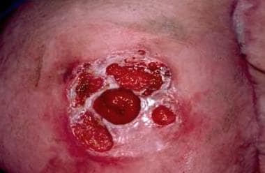
 Classic, or typical, pyoderma gangrenosum. This patient did not have an associated disease, and the condition responded well to cyclosporine.
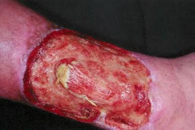