

Researchers at Aarhus University have used cryo-EM to gain a better understanding of the folding process that dictates RNA origami. Among other things, they found a kinetic phase from which an RNA structure could only free itself after ten hours, they write in Nature Nanotechnology.
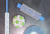
RNA is an excellent building block for creating diverse, well-defined structures at the nanoscale. The power of RNA origami lies precisely in the fact that the RNA strands eventually achieve the desired three-dimensional shape through self-assembly and programmed folding. For example, a neat little bundle of interconnected helices that together form a larger hollow cylinder in which a cargo can be transported. Or, conversely, a flat stack of helices that acts as a kind of ’wall’ that can be used to build ’scaffolds’ for applications in synthetic biology.
Design rules
Design ideas abound, but the big question with RNA origami, and the slightly more advanced field of DNA origami, is what sequence you need to get exactly the structure you want. You need clear design principles, design rules, so that you can proceed in a rational way. But finding these rules requires a very detailed understanding of the resulting RNA structures.
So far, these structures have been studied mainly using atomic force microscopy and transmission electron microscopy. The disadvantage of these techniques is that the RNA constructs are attached to a surface, which not only perturbs the structure, but also does not correspond to the final environment in which these structures fold and must function, namely the aqueous environment of biological tissue.
Freezing
Researchers at Aarhus University in Denmark therefore chose cryo-EM (electron microscopy with frozen samples) to study a set of common RNA origami structural elements at very high resolution. In this way, the RNA structures are in a much more natural conformation because they are folded in solution and not attached to anything.
‘A very interesting paper’, says Tom de Greef, Professor of Synthetic Biology at the TU Eindhoven. ’By using cryo-EM, the authors manage to characterise the structures much better, and by that I mean that the conditions are much closer to the real situation due to the interaction with water.’
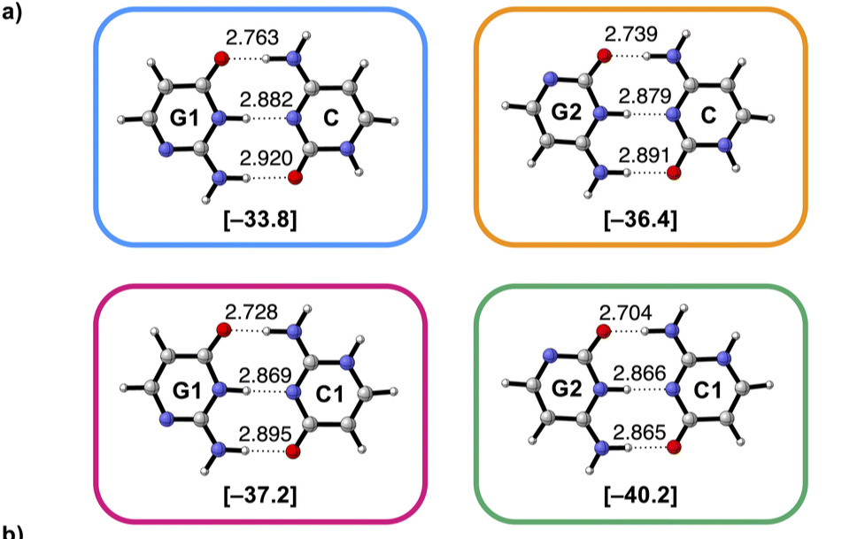
Among other things, an earlier assumption about the parallel arrangement of helices in a bundle turned out to be incorrect. The helices prefer to be at a small angle to each other. Another assumption, that kissing loops - in which the RNA strand forms a hairpin structure and sticks back to itself - always form a tight 180-degree bend and consist of 8-9 base pairs, was confirmed. A striking finding was the appearance of a kinetic folding trap in one of the designs. This is where a different structure than expected initially forms, and only after ten hours do the strands adopt the intended, thermodynamically most favourable structure.
The cycle of design - build - test - learn that the authors follow provides insight into the design rules, says De Greef. ’They discover important parameters for the formation of loops and kinetic traps during the folding process and show that they can modify the design to optimise these parameters and avoid the traps. The use of cryo-EM for structure determination is crucial. ’It shows that analysis using methods such as cryo-EM, where the RNA origami folds in its natural environment, i.e. in the presence of water, is very important to be able to optimise the formation of these structures.’
Ewan McRae, et al, Structure, folding and flexibility of co-transcriptional RNA origami, Nature Nanotechnology (2023)


Nog geen opmerkingen